About magnetic tomography
What is Magnetic tomography or MRI?
Magnetic tomography in Chisinau is a modern imaging diagnostic technique. It uses the combination of an intense magnetic field and radio waves with specific frequencies to visualize at a higher clarity and resolution the internal structures of the human body, making it possible to accurately diagnose many conditions.
Radio frequency signals, perceived by the patient as loud repeated noises, emitted by the device are captured and then retransmitted by the patient's body resonance phenomenon. Captured by special antennas fixed on the investigation area and taken by the computer to be transformed into multiplanar images like tomographic sections in any coordinate plane.
BENEFITS
- modern method of non-radiant, non-invasive investigation;
- does not pose a danger to the human body;
- offers possibilities for multiplanar representation;
- detects very small lesions (0.5 mm);
- recommended in investigations of all organs.
CONTRAINDICATIONS
- cardiac pacemaker;
- cochlear implants;
- electronic devices or implants;
- devices / implants with magnetic activation (hip prostheses, rods, metal heart valves or any other kind of metal fixed on the body;
- neurostimulators / medullary stimulators.
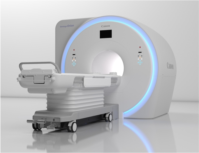
CANON TECHNOLOGY of 1.5 Tesla
MAGNETIC FIELD OF SUPERIOR HOMOGENEITY
the strength of the 1.5 Tesla magnetic field and the most sophisticated scanning applications offer the possibility of its use in diagnosing a very wide range of diseases producing high quality images.
EXCEPTIONAL IMAGE QUALITY
the Japanese production device CANON Vantage ORIAN is equipped with the most advanced technologies of signal processing and image reconstruction that are often comparable to MRIs with 3 Tesla magnetic fields - used for research purposes.
WIDEST OPENING - 71 CM IN DIAMETER
we have the largest aperture (hole) in the MRI industry - 71 cm which allows scanning of overweight patients.
DISPLAY OF VESSELS WITHOUT CONTRAST
we are equipped with the technology that allows the visualization of intra and extracranial vessels without the contrast substance - TOF protocol.
LOW NOISE LEVEL
special noise attenuation technology.
ADVANCED SCAN PROTOCOLS
We have protocols developed in scanning different anatomical parts of the body to diagnose different conditions and diseases that otherwise cannot be visualized by other methods and lower level MRI devices.
Our Services
Unique 1.5 Tesla magnetic resonance technologies
Services of magnetic tomography in Moldova with 1.5 Tesla magnetic field strength and the most sophisticated applications for scanning and reconstructing medical images offer the possibility of using it to diagnose a very broad spectrum of diseases while producing images of superior quality.
LARGEST APERTURE – 71 CM IN DIAMETER
MRI has the largest aperture (hole) in the industry – 71 cm in diameter, allowing it to scan overweight patients.
VISUALISATION OF VESSELS WITHOUT CONTRAST
Magnetic tomography is equipped with technology that allows visualization of intra- and extracranial vessels without the contrast agent with superior image quality - the TOF protocol.
LOW NOISE LEVEL
All devices produce noise during scanning which creates discomfort for patients. But the MRI in our equipment produces a much lower level of noise.
EXCEPTIONAL IMAGE QUALITY
Services of magnetic tomography in Chisinau from japanese-made device of Canon Medical Systems Corporation is equipped with the most advanced signal processing and image reconstruction technologies that are often comparable to 3 Tesla magnetic field MRIs - used for research purposes.
ADVANCED SCANNING PROTOCOLS
We have developed protocols for scanning different anatomical parts of the body to diagnose various conditions and diseases that cannot otherwise be visualized by other methods and lower-level MRI devices.
You can see the prices for magnetic tomography on the right!
Sign up for our other services of tomography – Computed tomography
MRI in Moldova – only CEIM.md!
-
MRI of brain standard protocol2 200 lei
-
MRI angiography of intracranial vessels MRA and MRV without contrast2 600 lei
-
MRI of brain standard protocol with contrast4 100 lei
-
MRI of pituitary gland without contrast (pituitary, turkish saddle)2 100 lei
-
MRI of the pituitary gland with contrast (pituitary, turkish saddle)4 100 lei
-
MRI of cervical region (soft tissues)2 400 lei
-
MRI angiography of intracranial vessels MRA without contrast2 400 lei
-
MRI of the cervical region (soft tissues) with contrast4 200 lei
-
MRI of spine 1 region (cervical, thoracic, lumbosacral, coccygeal)2 200 lei
-
MRI of spine 1 region with contrast4 100 lei
-
MRI of spine 2 regions (cervical, thoracic, lumbosacral, coccygeal)3 800 lei
-
MRI of spine 3 regions (cervical, thoracic, lumbosacral, coccygeal)5 400 lei
-
MRI of joint (shoulder, elbow, radiocarpal, knee, talocrural)2 200 lei
-
MRI of a joint (shoulder, elbow, radiocarpal, knee, talocrural) with contrast4 100 lei
-
MRI of hip joints2 200 lei
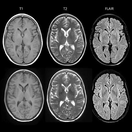
Brain MRI
Brain MRI - is the method of choice in the evaluation of brain structures, being useful in the assessment and monitoring of a wide range of pathologies:
✓ Tumour pathology - brain or meningeal tumours;
✓ Vascular pathology - stroke, arterio-congenital malformations, venous sinus thrombosis, aneurysms;
✓ Malformative pathology - congenital malformations of the brain, congenital malformations of the meninges;
✓ Degenerative pathology - epilepsy, internal hydrocephalus, cerebral atrophy,;
✓ Pathology of the orbit, optic nerves and eyeball muscles - inflammation or neuroendocrine disorders, diplopia, tumours;
✓ Pathology of the inner ear - inflammatory, tumoral, malformative, labyrinthine lesions, vascular abnormalities;
✓ Pathology of pituitary region - micro and macro pituitary adenoma, visualization of pituitary rod;
✓ Inflammatory pathology - encephalitis, meningitis, encephalopathies, leukodystrophies, multiple sclerosis;
✓ Brain pathology in systemic diseases - leukaemia, lymphoma, HIV-AIDS;
✓ Post-traumatic pathology.
In certain pathologies, in addition to a standard examination, special sequences are required, such as: MR angiography (arterial/venous), MR perfusion, DTI + tractography, MR spectroscopy.
preparation
General preparation for magnetic tomography investigation
Before the examination the patient may eat and take medication according to the daily schedule, unless otherwise specified. As the patient is under the infusion of a strong magnetic field, it is recommended that jewellery, watches, cards, hearing aids, dentures, hairpins and any other metal accessories are removed before the examination.
The patient lies on the examination table and is instructed to remain as still as possible during imaging. Tell the doctor if you have implants or medical devices in your body. Most of the time, MRI is safe for patients with new-generation metal implants, but patients with cochlear implants and almost all pacemaker wearers cannot have the investigation.
If contrast material injection is required, the patient should inform the radiologist if they suffer from any allergies (food, medication), or if they have asthma. It is also important to mention the presence of kidney disease.
Although there are no reports of adverse effects on pregnant women or unborn babies, the examination is not performed on women in the first 3-4 months of pregnancy except in limited cases.
Special preparation for abdominal MRI.
MRI of internal organs requires special preparation. The accuracy of the results depends on its quality, so treat the procedure with utmost responsibility!
- A few days before the procedure, exclude foods that cause flatulence from your diet: alcohol, carbonated water, dark bread, legumes, dairy products, raw fruit, vegetables. Also stop using medications that cause increased gas formation.
- To increase the informative value of MRI of the spleen, pancreas or liver, eliminate carbohydrates from the diet.
- If you have constipation or frequent flatulence, take a course of laxatives or carminative medication.
- On the eve of a renal MRI, have a blood and urine test.
- Visit the bathroom before the investigation. The investigation takes 30-40 minutes. You will not be able to go to the bathroom during the scan.
- Remove all hairpins, rings, chains, earrings. They will affect the quality of the images.
- Before the investigation, you can only eat light food. The last meal should be 6-8 hours before the scan.
Before the scan during the day, it is forbidden to drink tea, coffee, carbonated drinks. Last fluid intake is acceptable 4 hours before the scan. If it is not your first investigation, take the previous results. This will help the doctor to understand if there are improvements, worsening, if other pathologies have appeared.