About computed tomography
What is Computed Tomography (CT)?
Computed tomography in Chisinau (CT) is an It is an advanced method of non-invasive medical imaging, combining X-rays with computer technology to obtain clear and detailed images of the internal anatomical structures of the human body. The principle of obtaining information is totally different from magnetic resonance and consists in performing multiple, serial radiographs of the human body, which after a complicated mathematical calculation will make at the end of the examination a multiplanar volumetric reconstruction (in various planes) of the entire region of interest. It is used to investigate virtually every region of the human body, but is more accurate in assessing bones, internal organs, and blood vessels.
BENEFITS
- modern method of investigation;
- short lead time;
- offers possibilities for multiplanar representation;
- detects very small lesions (<0.5 mm);
- recommended in investigations of all organs.
CONTRAINDICATIONS
- pregnancy.
CONTRAINDICATIONS in case of investigations with contrast i / v
- the patient is hyperalergic to any medicine or even to the contrast substance;
- kidney failure, severe heart failure;
- pathologies of the thyroid gland.
Benefits:
+ AIDR 3D (Adaptive Iterative Dose Reduction 3D) - integrated software for automatic control of radiological exposure that allows maximum optimization of the irradiation dose depending on the patient's body mass, area of interest and scanning protocol.
+ SEMAR (Single Energy Metal Artifact Reduction) - complex algorithm that uses sophisticated reconstruction techniques to reduce metal-induced artifacts (dental implant, metal stents) and allows the analysis of adjacent soft tissues with increased accuracy.
+ DUAL ENERGY - is a fairly new technology that synchronously uses two different energies of exposure values (primary and secondary roentghen rays) to capture images. This unique regime allows flexibility in the post-processing of acquired images, therefore it can improve vascular imaging by increasing the attenuation of vessels at lower energies, providing an improved view of highly calcified vessels with the help of exposure dose reduction techniques. Another application of this technique is to determine the type of stones (urates, oxalates) in urolithiasis.
+ PEDIATRIC CARE - pediatric protocols adapted according to age and body weight, in accordance with world standards, allow the investigation of children with a minimum dose of exposure.
+ HEAVY CARE - the TC Aquilion Prime installation has the largest opening (hole) of 80 cm and the mass of the installation has a lifting capacity of up to 200 kg. In addition, the facility is equipped with specially adjusted scanning protocols for overweight people.
Our Services
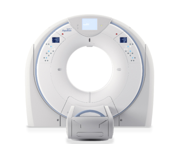
Unique technologies of computed tomography with 160 sections
Services of computed tomography in Moldova with 160-slice scanning and 35 millisecond rotation speed offers superior clinical benefits to all types of patients.
WE HAVE THE LOWEST DOSE OF TC IRRADIATION
The CT scanner is equipped with a unique technology that reduces the irradiation dose by up to 60% compared to other existing technologies.
LARGEST APERTURE - 78 CM IN DIAMETER
The Computed Tomography scanner has the largest aperture (hole) of 78cm - which allows scanning of overweight and claustrophobic patients.
CT SCANNING WITH IMPLANTS PRESENT
The Computed Tomography scanner reduces metal artefacts (dental implants, metal stents), allowing analysis of adjacent soft tissues with increased accuracy in patients with prostheses or pacemakers.
FAST CT SCANNING SPEED
Services of computed tomography in Chisinau are very fast. The tomographer makes a full rotation around the patient in just 0.35 seconds, which requires minimal withholding of resuscitation or precludes the need for it - an important consideration in scanning pediatric patients, and elderly or anxious patients.
DUAL ENERGY TECHNOLOGY
This unique scanning regime offers flexibility in post-processing of acquired images, therefore can improve vascular imaging by increasing vessel attenuation at lower energies, providing improved visualization of heavily calcified vessels using dose-lowering techniques.
SCANNING CHILDREN WITH LOW DOSES OF IRRADIATION
We have paediatric scanning protocols, tailored according to age and body mass, in line with global standards, allowing investigations to be performed in children with minimal exposure dose.
You can see the prices for computed tomography on our website!
Sign up for our other services of tomography – Magnetic tomography
MRI in Moldova – only CEIM.md!
-
CT of brain without contrast1 400 lei
-
CT of brain with contrast3 500 lei
-
CT of paranasal sinuses1 300 lei
-
CT of paranasal sinuses and brain1 700 lei
-
CT of nasopharynx or larynx1 400 lei
-
CT of spine 1 - anatomical region (cervical, thoracic, lumbar, sacral, coccygeal)1 400 lei
-
CT of chest and mediastinum without contrast1 400 lei
-
CT of chest and mediastinum with contrast3 500 lei
-
CT of abdomen without contrast1 400 lei
-
CT of abdomen with contrast3 500 lei
-
CT of small pelvis without contrast1 400 lei
-
СT of small pelvis with contrast3 500 lei
-
CT of joints 1 - anatomical region1 400 lei
-
CT of anatomical region (femur, calf, arm, forearm, neck)1 400 lei
-
CT of anatomical region (femur, calf, arm, forearm, neck) with contrast3 500 lei
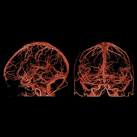
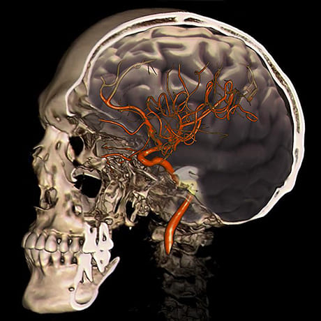
Cerebral CT angiography
Cerebral CT angiography - is an advanced diagnostic method that involves intravenous administration of contrast dye to visualise and assess cerebral arteries/veins. It is a highly informative investigation for the detection of aneurysms and arteriovenous malformations and tumours, as well as for planning their surgical treatment.
In the case of stroke, CT angiography is useful for determining the ischaemic or haemorrhagic aetiology of stroke. In the case of atherosclerosis of the magistral vessels it can differentiate between a total occlusive thrombus, subocclusive thrombus or atherosclerotic stenosis.
For the evaluation of acute stroke patients, CT angiography is a rapid and accurate method, with relevant information regarding both vascular caliber and patency and parenchymal perfusion being obtained at the first pass of bolus contrast medium.
preparation
General preparation for Computed Tomography
Preparing the patient for the CT scan is not a complicated process. If an investigation of the abdomen is to follow, it is recommended that the patient does not eat for 4 hours beforehand, and in other cases it may be necessary to take laxatives or perform an enema to thoroughly cleanse the lower intestinal tract before the CT scan.
If an examination in the head area follows, metal objects such as earrings or dentures should be removed so as not to affect the CT scan result.
Special preparation for contrast CT scanning
-
The patient must not eat any food on the day of the investigation
- Must submit results of urea and creatinine tests (not older than 10 days)
- Patients receiving treatment for diabetes mellitus should stop taking preparations 3 days before the date of the investigation
- If the patient has had previous investigations with contrast, they will present the results of the investigation on the day of the appointment.
Special preparation for cholangiographic MRI (MRCP)
- A few days before the procedure, exclude foods that cause flatulence from the diet: alcohol, carbonated water, dark bread, legumes, dairy products, raw fruit, vegetables. Also stop using medicines that cause increased gas formation.
- To increase the informative value of MRI of the spleen, pancreas or liver, eliminate carbohydrates from the diet.
- If you have constipation or frequent flatulence, take a course of laxatives or carminative medication.
- On the eve of a renal MRI, have a blood and urine test.
- Visit the bathroom before the investigation. The investigation takes 30-40 minutes. You will not be able to go to the bathroom during the scan.
- Remove all hairpins, rings, chains, earrings. They will affect the quality of the images.
- Before the investigation, you can only eat light food. The last meal should be 6-8 hours before the scan.
- On the day of the investigation, the patient does not eat anything and must drink a No Spa Forte pill ( 2-3 hours before the investigation). 30-60 min before the investigation the patient must drink 250 ml of pineapple juice (no other components, only pineapple).
Special preparation for MRI of the small pelvis with or without contrast
Patients requesting MRI of the small pelvis, the last day of the menstrual cycle must be specified (7-10 days after the last day of the menstrual cycle must pass in order to perform the investigation).